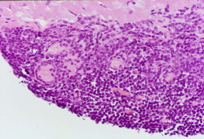
Figure 2.
A higher magnification of the Quilty lesion shown in
Figure 1, reveals the distinguishing characteristics. Note that the
infiltrate is composed almost exclusively of small lymphocytes,
which occasional macrophages and plasma cells. There are
also small vascular channels that have a prominent endothelium.
Please mail comments, corrections or suggestions to the
TPIS
administration at the UPMC.