PART B:
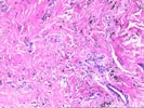
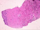
RIGHT LEG LESION, BIOPSY -
- CHANGES MOST CONSISTENT WITH STASIS DERMATITIS (see microscopic description and diagnostic comment).
Comment:
The differential diagnosis between stasis dermatitis and the
patch stage of Kaposi's sarcoma is a difficult one. At present,
we favor the diagnosis of stasis dermatitis, because of the
histopathological findings(see microscopic description) and the
clinical history of long standing stasis changes in the lower
extremities(as per Dr. Ragni). Additional information which may
be helpful in distinguishing between these two possibilities
include the detection of the Kaposi's sarcoma virus. If these
studies can be carried out on formalin-fixed paraffin-embedded
tissue, an addendum report will follow. Dr. Randall Lee also
reviewed this case and concurs with the interpretation.
Previous Biopsies on this Patient:
None
Gross Description - Case 1
The specimen consists of five (5) consult slides 97 with an accompanying surgical pathology report.Microscopic Description - Case 1


Slide A consists of skin with an acanthotic epidermis showing focal ulceration with underlying granulation tissue containing a severe acute and chronic inflammatory infiltrate, which consists predominantly of mature plasma cells. No definite viral inclusions or definite cytologic atypia is appreciated. The overlying epithelium shows pseudoepitheliomatous hyperplasia and hyperkeratosis with parakeratosis.
Section B also shows a section of skin with mildly acanthotic epithelium and some flattening of the rete ridges. However, the most striking changes are in the dermis where there is collagenization of the dermal connective tissue. This is intermixed with abundant deposition of pigment, which is apparently hemosiderin. Also present are small lobules of blood vessels which show Factor VIII positivity. Very little inflammatory cell infiltrate is seen. No definite insinuation of endothelial cells between collagen bundles are seen, and no clusters of spindled cells can be appreciated. Moreover, we do not see the characteristic pattern of larger dilated vessels and surrounding, smaller proliferating vessels. Instead, the increase in vascularity seems to be arranged in lobular aggregates, and there is abundant hemosiderin deposition. Based on these findings, a diagnosis of stasis dermatitis is favored over the patch stage of Kaposi's sarcoma at this time.
Please mail comments, corrections or suggestions to the TPIS administration at the UPMC.

