Previous Biopsies on this Patient:
None
TPIS Related Resources:
Liver Transplant Topics


(2 HE, 1 Trichrome)
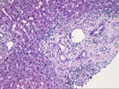
The normal lobular architecture is distorted by portal expansion because of duct and cholangiolar
proliferation and focal portal fibrosis. Native bile ducts are
intact and the normal spacial relationship between portal tracts
and central veins is maintained in mostly all areas. There is
also centrilobular hepatocanalicular cholestasis and a focal bile
infarct, indicative of biliary tract obstruction and stricturing.
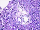
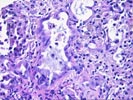
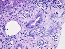
On closer examination, several of the expanded portal tracts and one large fibrous area contain markedly atypical, neoplastic- appearing epithelial cells, some of which are arranged into small irregularly shaped glands that show focal cribriforming. Cytologically, these atypical glands are composed of cells with large irregular and even some bizarre hyperchromatic nuclei, with irregular nuclear membranes and occasional cells contain large bizarre irregular eosinophilic nucleoli. There is a small to moderate amount of eosinophilic cytoplasm with focal mucin production. Several of the neoplastic cells contain intracytoplasmic vacuoles indicative of signet ring differentiation and occasional mitotic figures are also seen. The neoplastic glands and individual neoplastic cells are seen infiltrating an edematous, desmoplastic-type stroma and apparent angiolymphatic invasion is also seen.
Overall, the histopathological changes are consistent with an adenocarcinoma. This impression is based on the architectural arrangement and distribution and the cytological characteristics of the atypical cells. The apparent intrahepatic angiolymphatic invasion is most consistent with a biliary tract primary. This case was also reviewed at our quality assurance conference of 08- 12-97 and all present agreed with the interpretation.