 Contributed by Michael A. Nalesnik, M.D.
PATIENT HISTORY:
Per referral patient history, the patient is a 42-year-old male
with a chief complaint of bloating x 1 1/2 weeks. The patient
has a history of psoriasis and seizures complaining of occasional
lower abdominal pain and abdominal bloating beginning
approximately one and a half weeks ago. The patient reports a
decreased appetite for the same amount of time. Possible yellow
skin per patient's mother, times one week. Positive dark, red
urine, times one and a half weeks. Positive lower extremity
swelling to hips on both sides for about the same amount of time.
The patient denies any shortness of breath, nausea, vomiting,
fever, chills, or night sweats. The patient is on Dilantin, 200
mg po BID and occasional Advil. He is allergic to codeine and
penicillin. He smokes one pack per day x 24 years. The patient
admits to eight beers a day x two years, but has a drinking
history for the past 25 years. He has been in rehab once, not on
his own accord. He denies shakes or withdrawal. He has periods
of no ETOH. He denies IV drug use, but states he has used
marijuana, cocaine and LSD in his adolescence. Review of outside
material.
Final Diagnosis (Case 66)
LIVER, BIOPSY -
ACTIVE MICRONODULAR CIRRHOSIS WITH UNDERLYING STEATOHEPATITIS
(See comment).
COMMENT:
The histologic features suggest cirrhosis secondary to
steatohepatitis. A possible etiology of this patient's
steatohepatitis is alcohol use as documented in his clinical
history. Additional underlying biliary disease is less likely
but cannot be entirely ruled out and clinical evaluation would be
necessary to document this. The florid ductular proliferation
appears to be associated with the underlying liver injury and
does not appear to represent a neoplasm.
Previous Biopsies on this Patient:
None
TPIS Related Resources:
Liver Transplant Topics
Gross Description - Case 66
The specimen consists of one (1) consult slide and one (1)
paraffin block with an accompanying surgical pathology
report and patient history.
Microscopic Description - Case 66
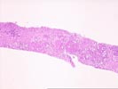
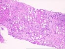
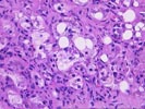
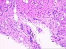
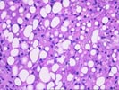
(1 HE)
The liver needle core biopsy shows distortion by nodules
surrounded by extensive fibrosis. The fibrous septae show mild
mixed inflammation consisting of neutrophils, lymphocytes, rare
histiocytes and rare plasma cells. The septae show extensive
cholangiolar proliferation. There is mild interface activity.
Cholestasis is present near the limiting plate. The lobules show
pericellular fibrosis and scattered neutrophils. These
neutrophils in some areas surround swollen degenerating
hepatocytes. Some of the swollen hepatocytes show Mallory's
hyaline. Some areas suggestive of scar like fibrosis are seen
within the fibrous septae.
Please mail comments, corrections or suggestions to the
TPIS administration at the UPMC.
)
)
)
)
)