Previous Biopsies on this Patient:
None
TPIS Related Resources:
Liver Transplant Topics

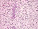
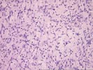
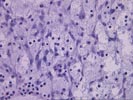
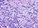
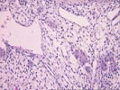

(4 HE) Sections reveal a fairly well-circumscribed mass, which appears to be fairly well separated from the surrounding hepatic parenchyma by fibrous tissue containing cholangioles. The portal triads in the surrounding "normal" liver show cholangiolar proliferation - an apparent mass effect.
The lesion consists predominantly of mesenchymal cells, which are embedded in a diffusely myxoid background, containing strands of mature hepatocytes and traversed by multiple venous, arterial, and capillary-like vascular channels. Cytologically, the stromal cells consist of non-descript, spindle-shaped cells with a moderate amount of eosinophilic cytoplasm. Foci of extramedullary hematopoiesis are also present. In one slide, mitotic activity can be seen in some of the stromal cells (2- 3/HPF). Ductular structures are not particularly prominent.
Overall, the histopathological changes and the clinical history are most consistent with a mesenchymal hamartoma, assuming that the immunostains mentioned in the history were all negative. However, an infantile hemangioendothelioma cannot be absolutely excluded. A Factor VIII related antigen and/or CD31 or CD34 stain may be helpful in differentiating between these two diagnostic possibilities. I took the opportunity of sharing this case with Dr. Ron Jaffe at Children's Hospital, who favored the diagnosis of infantile hemangioendothelioma and who sugested that other vascular lesions in the liver should be excluded. Dr. Randall Lee favored a diagnosis of mesenchymal hamartoma. I look forward to any follow-up on this case.