Previous Biopsies on this Patient:
None
TPIS Related Resources:
Knodell Scoring
Liver Transplant Topics

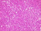
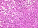
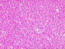
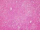
The sections show fragments of a benign hepatocellular neoplasm characterized by the absence of normal lobular landmarks, the presence of scattered aberrent arteries, and the proliferation of cytologically bland liver cells in plates of one to two cell thickness. Some fragments also show multiple efferent veins in abnormal relationships. The liver cells show mild macrovesicular steatosis, but no nuclear atypia, mitoses, or evidence of invasion are identified. These changes are consistent with a hepatic adenoma, and a clinical history of oral contraceptive or other steroid use would be of interest. No evidence of metastatic breast carcinoma is seen.