COMMENT:
No specific etiologic clues are identified, although the changes
are consistent with an underlying autoimmune hepatitis. Because
of the steatosis and foci of pericellular fibrosis, the
possibility of an inactive steatohepatitis should also be
considered.
Previous Biopsies on this Patient:
NONE
TPIS Related Resources:
Knodell Scoring
Liver Transplant Topics
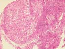
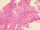
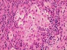
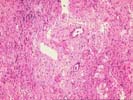
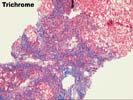
The liver biopsy is small and partially fragmented with a zone of subcapsular collapse. Nonetheless the architecture is deranged and deeper portions of the biopsy demonstrate significant fibrosis and architectural derangement consistent with established cirrhosis. There is a moderate mononuclear infiltrate within the fibrous tissue, which is associated with mild piecemeal necrosis. Occasional plasma cells are identified. The bile ducts appear intact and no florid duct lesions are identified. The nodules demonstrate moderate macrovesicular steatosis. Occasional swelling of the liver cells is seen, but definite Mallory's bodies are not identified. The trichrome stain identified bridging fibrosis and additionally cirrhosed zones of pericellular fibrosis. The iron stain is negative. No viral inclusions, ground glass hepatocytes, pigment deposition or cytoplasmic globules are identified.