Previous Biopsies on this Patient:
NONE
TPIS Related Resources:
Knodell Scoring
Liver Transplant Topics
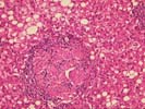
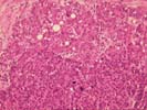
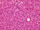
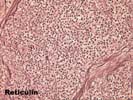
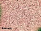
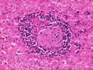
Sections show a liver cell neoplasm, which is growing in trabeculae and small sheets, separated by relatively thin bands of lamellar-type fibrosis in areas. The center of the mass contains large blood vessels, some of which appear to have previously undergone thrombosis.
On closer examination, some of the neoplastic cells form pseudoacini. Bile production is focally seen as are apparent pale bodies, mild steatotic change, and Mallory's-like hyaline. One small portal vein in Section BF contains several neoplastic cells near a small granuloma, which appear to extend into a small portal vein branch. However, unequivocal vascular invasion is not identified. At the interface between the tumor and the surrounding liver, the neoplastic cells irregularly extend into the uninvolved non-cirrhotic parenchyma.
Cytologically, some of the cells are quite large, with a high N:C ratio, abundant granular eosinophilic cytoplasm, large hyperchromatic irregular nuclei and prominent eosinophilic nucleoli. In other areas, the tumor is composed of relatively small cells with a high N:C ratio growing in a sheet-like pattern, with focal disruption of the reticulin architecture. Mitotic activity varies throughout the tumor: in the most active regions there is approximately 1 mitosis/HPF.
Overall, the histopathological changes are diagnostic of a moderately well-differentiated hepatocellular carcinoma. The diagnosis is based on the overall architectural pattern, including focal disruption of the reticulin framework and irregular extension into the surrounding liver; the cytological changes in many of the cells, including the large hyperchromatic and irregular nuclei; and the increased mitotic rate. The non- cirrhotic liver, the focal areas of lamellar fibrosis, the apparent pale bodies, large eosinophilic cells with large hyperchromatic nuclei and prominent eosinophilic nuclei raise some suggestion of fibrolamellar differentiation. However, there are also many of areas of normal variant, moderately well- differentiated hepatocellular carcinoma, which is the preferred classification of this lesion.
The portal tracts and parenchyma of the uninvolved liver show a mass effect with mild, predominantly mononuclear inflammation and some contain non-caseating granulomas that do not impinge on the bile ducts. The same non-caseating granulomas are present in the tumor. Although it is not unreasonable to assume that the granulomas are a reaction to the tumor, a co-existent chronic granulomatous disease, such as sarcoidosis or deep fungal infection should be excluded. Clinical correlation and examination of special stains for microorganisms are suggested.