COMMENT:
The size, associated fibrosis, and multinodularity of the granulomas suggest the possibility of sarcoidosis. This possibility needs to be correlated with clinical and radiographic studies. Other differential considerations include infectious and drug-induced causes of granulomas. In a large minority of cases, however, no specific cause can be identified.
Previous Biopsies on this Patient:
NONE
TPIS Related Resources:
Knodell Scoring
Liver Transplant Topics
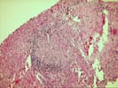
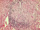
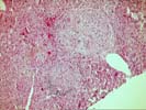
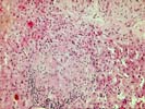
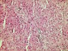
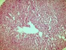
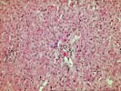
The liver biopsy shows intact hepatic architecture. There are several irregular foci of sinusoidal dilatation. The most striking finding is the presence of scattered well-formed epithelioid granulomas. These granulomas are present both within the lobules and within portal tracts, and they are associated with peripheral and segmentalizing fibrosis as well as, in some instances, a peripheral cuff of mononuclear cells. No bile duct loss or other features of primary biliary cirrhosis is seen. Otherwise, mild nuclear glycogenation of liver cell nuclei is identified. There is no significant hepatitis or other abnormality. The sinusoidal dilatation is presumably of local effect, resulting from the presence of granulomas.
The causes of hepatic granulomas are legion. Nonetheless, several features of this case suggest the possibility of sarcoidosis, and this should be further investigated. Features of primary biliary cirrhosis are not seen, but other differential considerations would include infectious and drug-induced granulomatous reactions. I understand that microbial stains are negative in this case. The extent to which this granuloma should be worked further will depend largely on the clinical findings.