 |
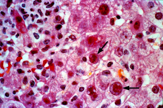
| Figure 3. A high magnification of the Adenoviral lesion shows the light microscopic appearance of the typical Adenoviral nuclear inclusions. Note the "smudgy" appearance of the nucleus(arrows), the peripheralized chromatin pattern, and the lack of cytoplasmic inclusions that help differentiate these inclusions from those seen with CMV. |