 |
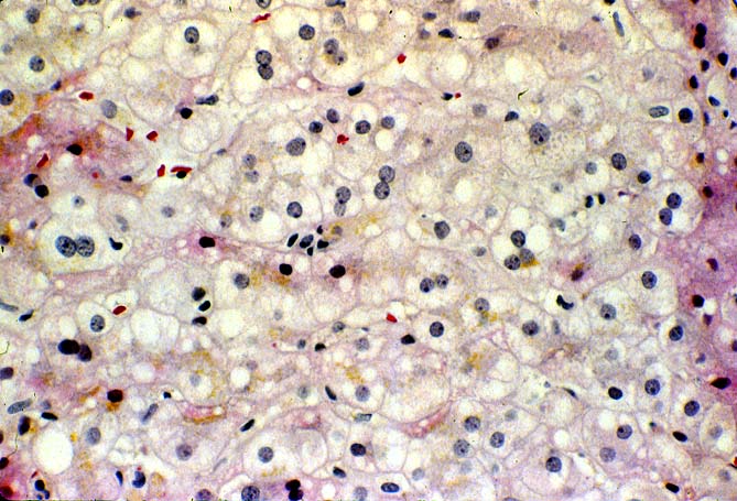
| Figure 2. In this photomicrograph there is low grade mononuclear lobular inflammation, typical of EBV-related hepatitis, like that seen in the general population with hepatitis related to infectious mononucleosis. Note the slight increase in sinusoidal lymphocytes and "indian-filing" along the sinusoids. This histopathological presentation is extremely difficult to differentiate from non-specific viral hepatitis or low grade viral hepatitis type B or C in an allograft recipient. The diagnosis of EBV hepatitis can be confirmed or excluded with the use of in situ hybridization for EBV EBER RNA. |