 |
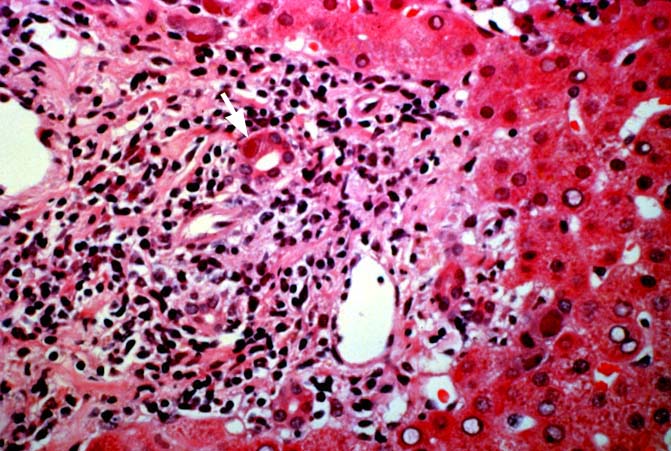
| Figure 9. This is a higher magnification of the portal tract and surrounding periportal hepatic parenchyma. Note the cytomegalovirus inclusion body present in the enlarged biliary epithelial cell [arrow]. Note also the lymphoplasmacytic infiltrate present in the portal tract, which can mimic acute cellular rejection if one misses the inclusion bodies. |