Approach to Tissue Specimens

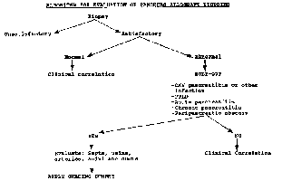
Post-Transplant Needle Biopsies

Workup for allograft dysfunction:

The major cause of graft loss with pancreas transplants is irreversible rejection. This is particularly true for pancreas after kidney (PAK) and pancreas transplant alone (PTA) cases since the clinical diagnosis of rejection remains relatively non-specific. When a simultaneous kidney transplant (SPK) is performed in an uremic diabetic the co-transplanted kidney is thought to provide a reliable indicator for rejection through serial determinations of the recipient's serum creatinine. Parameters used for the diagnosis of pancreas rejection include: decrease in urinary insulin and C peptide, increase in serum amylase, lipase, anodal trypsinogen and pancreas specific protein). Cytologic evaluation of pancreatic juice and urine have been also used in the diagnosis of pancreas allograft rejection. In bladder drained grafts, urinary amylase has been used as a measure of pancreas exocrine function. Other methods used for the diagnosis of pancreas rejection are 99mTc DTPA scintigraphy and uptake of indium-labeled platelets.

The success of renal, hepatic and cardiac transplantation, has been dependent on the ability to differentiate non-immunologic causes for graft dysfunction from rejection by reliance on a confirmatory biopsy. Biopsy material from transplanted pancreas was obtained in the past during laparotomy or through cystoscopically guided transduodenal pancreatic biopsy. The use of percutaneous pancreas graft biopsies has has yielded tissue for histologic analysis in greater than 88% of attempts, with complications in fewer than 2% of cases.

The biopsies are performed under ultrasound guidance using an 18-gauge automated biopsy needle with a 17mm specimen notch. The material is routinely fixed an processed. Twelve 4 micron sections are cut and levels 1,4,8 and 11 are stained with H&E. The remaining slides are used for additional stains if necessary (CMV, lymphoid markers, EBER, LMP-1, etc.) Or stored. The average surface area of each tissue section is 2mm3. Two to three segments of parenchymal lobules with corresponding fibrous septa are suitable for evaluation.

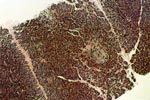
Protocol biopsies (normal pancreas, Grade 0):

Pancreas needle biopsies from patients with normal graft function typically show no inflammation. The reticulin stain highlights a delicate reticulin network outlining the acini and islets. Sometimes the tissue core includes parts of the peripancreatic fibrous tissue and skeletal muscle. In early post-transplant biopsies fat necrosis of the peripancreatic fat is common. The peripancreatic fibrous tissues often show foreign body giant cell reaction. Occasional biopsies contain fragments of benign lymph nodes. Biopsies from patients with normal function may also show inflammation of undetermined significance (Grade I).

REFERENCES

  1. Drachenberg CB, Papadimitriou JC, Klassen DK, et.al: Evaluation of pancreas transplant needle biopsy. Reproducibility and revision of histologic grading system. Transplantation 1997;63(11):1579-1586.
  2. Drachenberg C, Klassen D, Bartlett S, Hoehn-Saric E, Schweitzer E, Johnson L, Weir J and Papadimitriou J: Histologic grading of pancreas acute allograft rejection in percutaneous needle biopsies. Transplant Proc 1996;28(1):512-513.
  3. Drachenberg CB and Papadimitriou JC: Diagnosis of pancreas allograft rejection in percutaneous needle biopsies from patients with non-immune related graft pathology. Pathol Int 1996;46 (Suppl 1):533.
  4. Stratta RJ,Sollinger HW, Perlman SB et al: Early diagnosis and treatment of pancreas allograft rejection. Transplant Int 1988;1(1):6-12.
  5. Klassen DK, Hoehn-Saric EW, Weir MR, et al: Isolated pancreas rejection in combined kidney pancreas transplantation. Transplantation 1996;61(6):974-7.
  6. Sutherland DER, Casanova D and Sibley RK: Monitoring and diagnosis of rejection. Role of pancreas graft biopsies in the diagnosis and treatment of rejection after pancreas transplantation. Transplantation Proc 1987;19(1 pt 3):2329-2331.
  7. Jamieson C, Burry KD, Colapinto ND: Evaluation of intraoperative biopsy of the pancreas. Can J Surg 1977;20(6):546-550.
  8. Carpenter HA, Engen DE, Munn SR, et al: Histologic diagnosis of rejection by using cystoscopically directed needle biopsy specimens from dysfunctional pancreatoduodenal allografts with exocrine drainage into the bladder. Am J Surg Pathol 1990;14(9):837-846.
  9. Perkins JD, Munn SR, Marsh CL, Barr D, Engen DF and Carpenter HA: Safety and efficacy of cystoscopically directed biopsy in pancreas transplantation. Transplant Proc 1990;22(2):665-666.
  10. Allen RDM, Wilson TG, Grierson JM, Greenberg ML, Earl MJ, Nankivell BJ, Pearl TA and Chapman JR: Percutaneous biopsy of bladder-drained pancreas transplants. Transplantation 1991;51(6):1213-1216.
  11. Gaber AO, Gaber LW, Shokouh-Amiri MH, Hathaway D. Percutaneous biopsy of pancreas transplants. Transplantation 1992;54:548-550.
  12. Nakhleh RE and Sutherland DER: Pancreas rejection. Significance of histopathologic findings with implications for classification of rejection. Am J Surg Pathol 1992;16(11):1098-1107.
  13. Bartlett ST, Schweitzer EJ, Johnson LB, et al: Equivalent success of simultaneous pancreas kidney and solitary pancreas transplantation. A prospective trial of tacrolimus immunosuppression with percutaneous biopsy. Ann Surg 1996;224(4):440-449
  14. Drachenberg CB and Papadimitriou JC: Diagnosis of pancreas allograft rejection in percutaneous needle biopsies from patients with non-immune related graft pathology. Pathol Int 1996;46 (Suppl 1):533.
  15. Drachenberg CB, Papadimitriou JC, Weir MR, et al: Histologic findings in islets of whole pancreas allografts. Transplantation 1996;62(12):1770-2.


Underwritten By
Astellas

Please mail comments, corrections or suggestions to the TPIS administration at the UPMC.
Last Modified: Thu Jun 18 10:14:08 EDT 2009


Please give your feedback about this page here:


If you have more questions, you can always email TPIS Administration.