Virtually all patients who are infected with the hepatitis B virus(with
or without delta co-infection) and show evidence of viral replication
before transplantation(i.e., HBsAg or HBeAg sero-positive or HBV
DNA positive) will re-infect their allograft and develop hepatitis
after transplantation(1-15). Allograft
re-infection and thus, disease recurrence, is less predictable
in patients who had HBV-induced fulminant liver failure or in
those with chronic liver disease who had become anti-HBe positive
and serum HBV DNA and HBeAg negative prior to transplantation(1-12). Only 10-25% of this subpopulation
of recipients will not "re-infect" the allograft, nor
will they develop HBV-related disease after transplantation.
Unfortunately, despite donor and blood product screening, a small
number of patients without previous HBV disease will acquire HBV
infection during or after transplantation. This is noticeably
more common in recipients of HBcAb(+),
HBIgM(-), HBsAg(-) donors(16).
The highly infectious nature of HBV and the presence of extrahepatic
reservoirs of virus probably account for the difficulty eradicating
the virus before transplantation and its high recurrence rate
after transplantation. Nevertheless, various treatment modalities
have been used in an attempt to break this disheartening cycle
of re-infection and recurrent disease, which all too often, leads
to allograft failure and/or death. These include hepatitis B
immune globulin, various anti-viral drugs(17),
active vaccination with HBsAg, alpha-interferon (8, 18, 19), human anti-HBs monoclonal
antibody and the use of baboon liver donors for transplantation,
which are resistant to type B viral hepatitis infection(20, 21). Except for xenotransplantation(21), which is not yet a viable treatment alternative for
most patients, none of the above therapies can prevent re-infection.
However, continuous high dose anti-HBs therapy has been able
to delay the onset and ameliorate the injury associated with recurrence
of the B virus after transplantation(11, 14, 18, 22-26).
Since significant recurrent disease is a problem for most HBV+ recipients,
several programs questioned even whether liver transplantation
should be carried out in patients who are infected with the B
virus(8, 9, 27, 28). HBV+ recipients also have a high incidence
of complicating hepatocellular carcinomas that have the potential
to contribute to diminished patient and graft survival after transplantation(8, 29). Detailed studies however,
show that recurrence of the viral infection, and not the cancer, is the
major cause of morbidity and mortality after transplantation in HBV+ patients with co-existent hepatocellular carcinomas(8, 30). Moreover, encouraging reports
of the ameliorating effect of specific anti-viral therapy on disease
progression after transplantation have tempered the pessimism
such that many centers continue to offer liver transplantation
to patients infected with the B virus.
Pathophysiology
It is not our intention to engage in a detailed discussion of the
pathophysiology of hepatitis B virus-induced
liver disease. However, an overview of some of the pathophysiologic mechanisms of HBV-induced liver disease may help to explain some of the histopathological manifestations of HBV in an allograft liver.
Under normal circumstances, the hepatitis B virus is not thought to be cytopathic:
liver damage is thought to be at least partially attributable
to the expression of hepatitis B core antigen on the surface of
hepatocytes, which then become targeted for destruction by MHC-restricted
cytotoxic T lymphocytes (CTL)(31, 32). Thus, expression of viral antigens on the surface of hepatocytes precipitates lymphohistiocytic lobular lobular inflammation and spotty acidophilic necrosis of hepatocytes, typical of acute hepatitis. IN the general population, this is followed by "clearance" or control of the virus in most instances, or evolution toward chronic disease in a minority of recipients. In a liver allograft recipient, potent immunosuppression needed to prevent rejection interferes with normal mechanisms of viral clearing and contributes to the progression to chronic disease in most recipients. In fact, lower maintenance doses of corticosteroids may modify the course of post-transplant HBV infection by leading to less
viral replication, milder HBV-related liver disease and better overall patient survival(33). In addition, since there is no attempt to prospectively match the donor and recipient for MHC antigens, normal pathways of CTL lysis of virally infected hepatocytes may also be disrupted. Instead, viral could be processed by recipient accessory cells and presented to T lymphocytes in a MHC-class II restricted fashion, resembling a delayed type hypersensitivity response(3, 34).
Although MHC mismatching has the potential to significantly disrupt the manifestations of HBV-related disease in a liver allograft, there are too few cases at this time to draw any definite conclusions about how this actually occurs. Nevertheless, massive viral replication in class II MHC mismatched deteriorating liver allografts with little or no hepatic inflammation, prompted several groups, including our own, to suggest that, under special circumstances, the virus may be directly cytopathic(3, 5-7, 35, 36).
Clinical Presentation
HBV hepatitis usually first appears between six to eight weeks after
transplantation. Because liver injury tests are routinely monitored in this population, an elevation of the ALT and AST usually first prompts suspicion of recurrent disease. However, more dramatic initial presentations include nausea, vomiting, jaundice and hepatic failure, similar to that seen in the general population or in other immunosuppressed patients(1-12). The diagnosis is suspected on clinical grounds and confirmed after examination of a core needle biopsy.
Histopathologic Findings
Although local treatment policies can influence the
timing and severity of the histopathologic changes, in general, HBV hepatitis in liver allografts
is similar to that seen in non-allograft livers(1-10, 12, 35, 36). There is a typical progression from an acute hepatitis with predominantly lobular changes to chronic hepatitis, when a combination of portal tract and lobular changes are observed. Cirrhosis can develop with striking rapidity(1-10, 12, 35, 36). However, an occasional patient will
show histopathologic resolution of disease activity after a bout
of acute hepatitis, and rare patients will actually "clear"
the virus after transplantation. There also are several somewhat unique histopathological
presentations of HBV in the liver allograft recipient, that are likely
related to the potent immunosuppression and MHC non-identity between
the liver and recipient(see below).
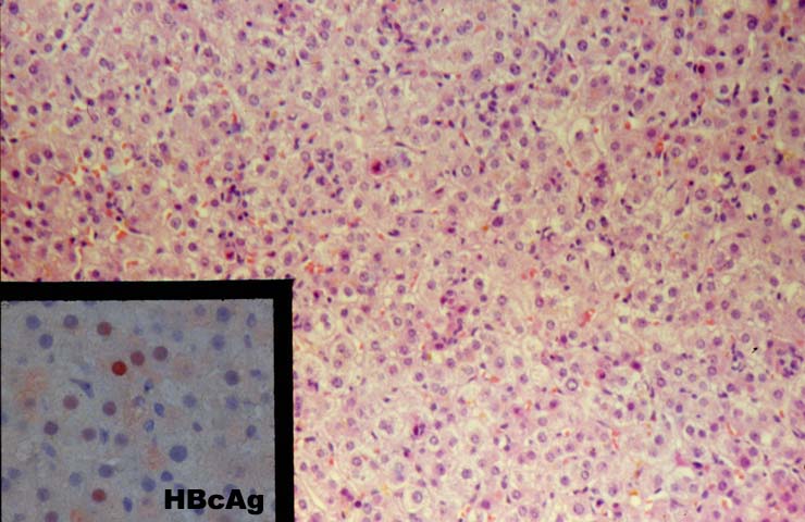
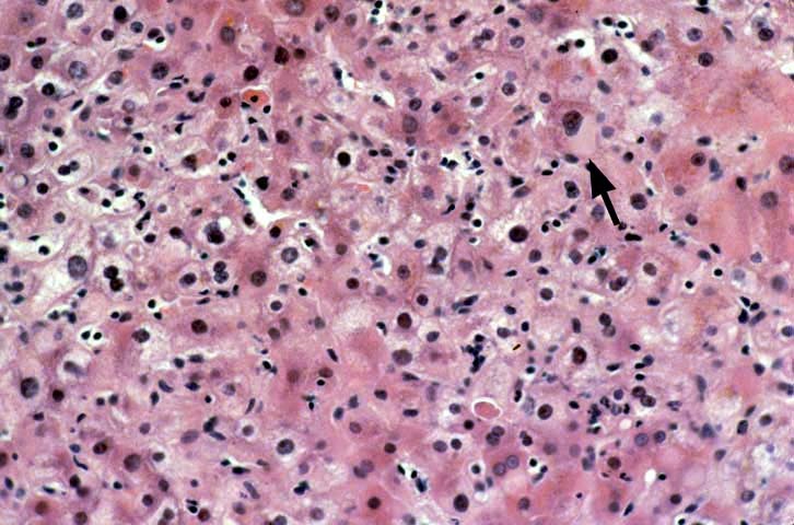
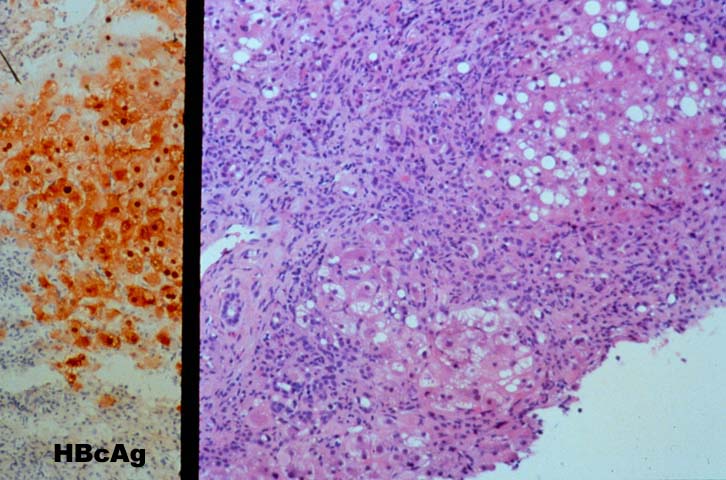
Re-infection with the type B hepatitis virus is usually first detected on immunohistochemical stains for HBV antigens. Within several weeks after transplantation, hepatitis B core antigen can appear in the cytoplasm of an occasional hepatocyte. The infection, and thus core and surface antigens,
spread throughout the liver lobules to involve a larger number of cells(1-10, 12, 35, 36). This is followed by acute hepatitis in which the lobular changes predominate. These include lobular disarray, Kupffer's cell hypertrophy, inflammation and necro-inflammatory activity; and variable portal inflammation. At this stage of the disease, a small percentage of patients will quickly progress to bridging or even submassive necrosis, particularly if immunosuppression is withdrawn(1). However, most patients show some resolution of the acute lobular necro-inflammatory activity, but evolution toward chronic hepatitis. The later phase is marked by predominantly mononuclear portal inflammation and interface activity. The lobular changes become less pronounced.
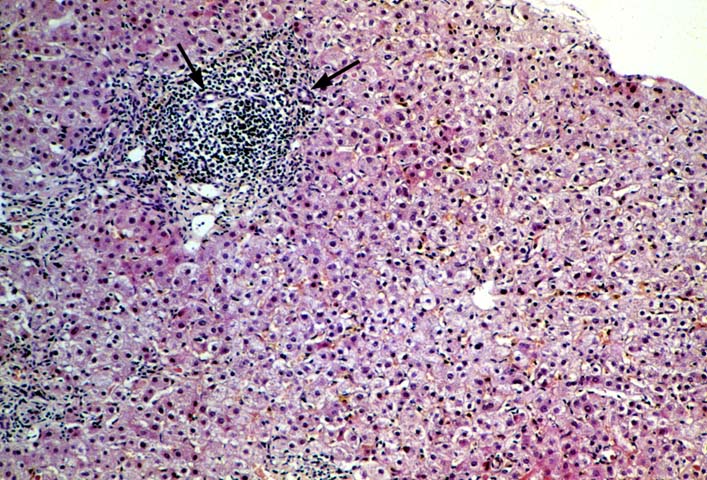
The typical case of chronic hepatitis in a liver allograft is similar to that seen in the general population and the same histological findings are used to ascertain the diagnosis. This includes lymphoplasmacytic portal inflammation that spares the bile ducts and portal veins, but shows
interface activity or peri-portal hepatitis of varying severity, characterized by extension of lymphocytes and macrophages into the edge of the lobule combined with cholangiolar proliferation. Lobular findings seen during the chronic phase of the disease include a large number of cells with a
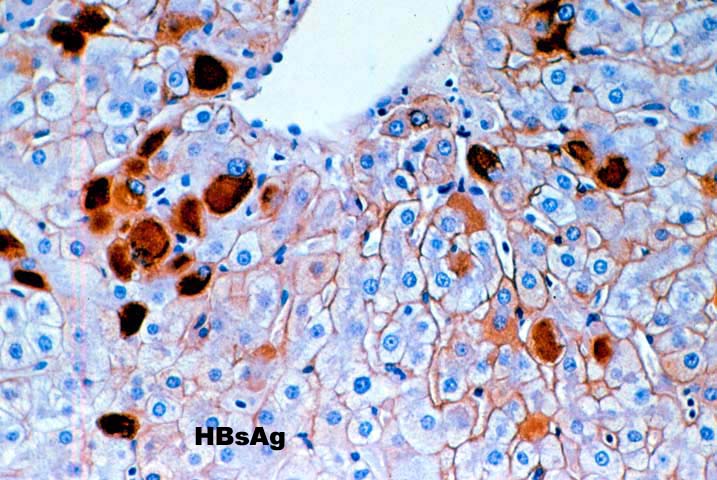
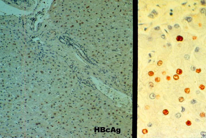
ground glass cytoplasm containing
hepatitis B surface antigen and, on occasion, numerous hepatocytes with sanded nuclei filled with
hepatitis B core antigen. This is accompanied by varying degrees of lobular disarray, regeneration, Kupffer's cell hypertrophy and lobular necro-inflammatory activity.
Unique Histopathological Presentations
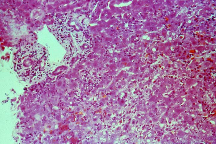
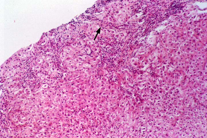
Several histopathological patterns of liver injury associated with HBV infection of a liver allograft are not commonly encountered in the general population. This is probably (but not definitely) related to effects of immunosuppression and MHC non-identity between the liver and recipient(3, 5-7, 10, 35, 36). The first unique histopathological pattern(3), termed "fibrosing cholestatic hepatitis (6)" is characterized by marked hepatocyte swelling, lobular
disarray and cholestasis, combined with minimal or no portal or lobular
inflammation, and cholangiolar proliferation of varying severity. Such cases are usually marked by
massive hepatocellular expression of core and/or surface antigen, that apparently lead to the hepatocyte
degenerative changes including cell swelling, steatosis and necrosis. Follow-up biopsies in such patients will often show progressive portal expansion because of cholangiolar proliferation, peri-portal sinusoidal fibrosis and lobular collapse, often without a significant inflammatory component(3, 5-7, 10, 35, 36). High levels of viral replication and antigen expression in these cases have led several groups to suggest that HBV may be directly cytopathic(3, 5-7, 10, 35, 36). Other less popular names that have been used to describe these constellation of findings, or ones very similar, include "fibrosing cytolytic hepatitis"(36) and "steatoviral" and "steatofibroviral" hepatitis (35) to emphasize the presence of hepatocellular cytolysis, steatosis and fibrosis, respectively.
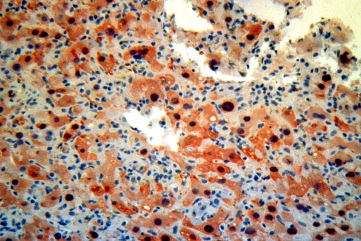
Finally, a massive overproduction of the surface antigen can be
seen in some patients. In these cases, the vast majority of hepatocytes contain a ground
glass cytoplasm, similar to HBsAg transgenic mice(37). This results in marked distortion of the hepatocytes, lobular disarray and mild necro-inflammatory lobular activity.
HCV and Delta Co-infection
In patients who undergo liver replacement for simultaneous HCV
and HBV disease, the recurrent hepatitis after transplantation more frequently resembled recurrent HBV than recurrent HCV. (38). In addition, one group suggests that the presence of HCV may actually improve the clinical outcome of recurrent HBV as compared HBV infection alone (39).
As in the general population, co-infection with the delta agent can complicate
HBV disease in the liver allograft recipient. The consequences of delta agent co-infection are not entirely clear because of somewhat conflicting follow-up results. There have been reports of both more and less severe disease after transplantation(10, 12, 40-43) compared to recipients with HBV alone. This might be related to the cytopathic effect of HDV in liver allograft recipients in relation to HBV replication. David et al(44) have noted that in patients with non-replicative HBV infection, HDV hepatitis produced histopathological lesions similar to those described in fibrosing cholestatic hepatitis. However, prominent HBcAg expression was not seen in the hepatocytes. In contrast, when HBV is actively replicating, co-infection with HDV resulted in necro-inflammatory activity similar to that seen in B & D viral hepatitis in patients from the general population(44).
Differential Diagnosis
During the acute phase, viral hepatitis type B should be distinguished from acute hepatitis caused by other "hepatitic" viruses such as type C and from "opportunistic viruses in the Herpes group. All can show a similar histopathological pattern of injury which includes lobular disarray, Kupffer's cell hypertrophy, lymphohistiocytic lobular inflammation, spotty acidophilic necrosis of hepatocytes and variable portal inflammation. The presence of inclusion bodies or ground glass cells can be used to distinguish HBV from hepatitis caused by viruses in the Herpes group. However, ground glass cells are uncommon during the initial presentation. Therefore, the distinction is usually made with the aid of special studies to detect viral antigens and/or nucleic acids in the blood or tissues.
At times, it can also be difficult to distinguish acute rejection from acute hepatitis since both can contain a mononuclear portal infiltrate(1, 3). However, the primary focus of the necro-inflammatory activity in rejection is distinct from that associated with viral hepatitis. In acute hepatitis, the lobule is the focus of injury, which is manifest as spotty acidophilic necrosis of hepatocytes, lobular disarray. Although varying degrees of portal inflammation can be seen, significant damage to portal
tract structures such as the bile ducts and veins is unusual. In contrast, the portal structures(portal vein and
bile ducts) are the focus of immunologically mediated damage in acute rejection (1, 3).
It can be extremely difficult to distinguish between late
onset acute or early chronic rejection from chronic hepatitis with low grade interface activity, particularly when no grand glass hepatocytes or sanded nuclei are seen. In such cases, a survey is taken of the percentage of damaged bile ducts. When there is damage of only an occasional bile duct and many bile ducts are un-inflamed, the cause of injury is more likely to be hepatitis. In contrast, rejection is more likely the cause of injury when there is damage of more than an occasional bile duct and a majority of the bile duct show degenerative changes such as eosinophilic transformation of the cytoplasm, uneven nuclear spacing and there is focal bile duct loss. In occasional cases it will be impossible to confidently distinguish between low grade acute rejection and chronic hepatitis on the basis of histopathology alone (45, 46). In such cases, our policy is to administer a bolus of corticosteroids and then follow the response to treatment over the next month. Typically, patients with rejection will show a more sustained response whereas those with viral hepatitis will transiently improve, but then relapse, with re-elevation of the liver injury tests after a period of a week or two. In some cases, rejection will primarily manifest as mononuclear inflammation in and around the terminal hepatic venules, which is often accompanied by perivenular hepatocyte dropout and sinusoidal red blood cell congestion. Even if the portal tracts show changes typical of hepatitis, in our experience, this so called "central venulitis" responds to increased immunosuppression (47).
Immunoperoxidase staining for HBV antigens is routinely carried out in almost all liver allograft biopsies from HBsAg+ recipients. In our experience, polyclonal antibody reagents are more sensitive but less specific than monoclonal anti-HBV reagents. Patients showing cytoplasmic HBcAg expression have experienced a more aggressive course in our experience (unpublished observation). However, detection of viral antigens does not mean that the liver inflammation and damage is due to the B virus(3). The final diagnosis should be based on the histopathological findings, including the pattern of injury.
REFERENCES
Demetris AJ, Jaffe R, Sheahan DG, et al. Recurrent hepatitis B in liver allograft recipients. Differentiation between viral hepatitis B and rejection. Am J Pathol 1986;125(1):161-172.
Portmann B, O'Grady J, Williams R. Disease recurrence following orthotopic liver transplantation. Transplant Proc 1986;18(4 Pt 5):136-141.
Demetris AJ, Todo S, Van Thiel DH, et al. Evolution of hepatitis B virus liver disease after hepatic replacement. Practical and theoretical considerations. Am J Pathol 1990;137(3):667-676.
O'Grady JG, Smith HM, Davies SE, et al. Hepatitis B virus reinfection after orthotopic liver transplantation. serological and clinical implications. J Hepatol 1992;14:104-111.
Davies SE, Portmann BC, O'Grady JG, et al. Hepatic histological findings after transplantation for chronic hepatitis B virus infection, including a unique pattern of fibrosing cholestatic hepatitis. Hepatology 1991;13(1):150-157.
Mason AL, Wick M, White HM, et al. Increased hepatocyte expression of hepatitis B virus transcription in patients with features of fibrosing cholestatic hepatitis. Gastroenterology 1993;105:237-244.
Todo S, Demetris AJ, Van Thiel D, et al. Orthotopic liver transplantation for patients with hepatitis B virus-related liver disease [see comments]. Hepatology 1991;13(4):619-626.
Lake JR, Wright TL. Liver transplantation for patients with hepatitis B: What have we learned from our results? [Editorial]. Hepatology 1991;13(4):796-799.
Lauchart W, Muller R, Pichlmayr R. Long-term immunoprophylaxis of hepatitis B virus reinfection in recipients of human liver allografts. Transplant Proc 1987;19(5):4051-4053.
Rizzetto M, Chiaberge E, Negro F, et al. Liver transplantation in hepatitis delta virus disease. Lancet 1987;2(8557):469-471.
Jansen PL, Haagsma EB, Klompmaker IJ, et al. - Hepatitis B-associated liver cirrhosis as an indication for liver transplantation. [Review]. Scand J Gastroenterol Suppl 1995;212:19-22.
Fung JJ, Eghtesad B, Todo S, et al. - Hepatitis B virus (HBV) re-infection following liver transplantation: theory and practice. [Review]. Clin Transplant 1995;9(3 Pt 2):262-268.
Wachs ME, Amend WJ, Ascher NL, et al. - The risk of transmission of hepatitis B from HBsAg(-), HBcAb(+), HBIgM(-) organ donors. Transplantation 1995;59(2):230-234.
Jamal H, Regenstein F, Farr G, et al. - Prolonged survival in fibrosing cholestatic hepatitis with long-term ganciclovir therapy. Am J Gastroenterol 1996;91(5):1027-1030.
Ranki M, Schatzl HM, Zachoval R, et al. - Quantification of hepatitis B virus DNA over a wide range from serum for studying viral replicative activity in response to treatment and in recurrent infection. Hepatology 1995;21(6):1492-1499.
Nour B, Tzakis A, Van Thiel DH. - The use of interferon for the treatment of viral hepatitis in pediatric liver transplant recipients [see comments]. J Okla State Med Assoc 1995;88(3):109-113.
Michaels MG, Lanford R, Demetris AJ, et al. - Lack of susceptibility of baboons to infection with hepatitis B virus. Transplantation 1996;61(3):350-351.
Starzl TE, Fung J, Tzakis A, et al. Baboon-to-human liver transplantation [see comments]. Lancet 1993;341(8837):65-71.
Ilan Y, Eid A, Tur-Kaspa R. - Long-term prevention of recurrent hepatitis B virus after liver transplantation. Isr J Med Sci 1995;31(8):469-473.
Langrehr JM, Lemmens HP, Keck H, et al. - Liver transplantation in hepatitis B surface antigen positive patients with postoperative long-term immunoprophylaxis. Transplant Proc 1995;27(1):1215-1216.
Samuel D, Bismuth A, Serres C, et al. HBV infection after liver transplantation in HBsAg positive patients: Experience with long-term immunoprophylaxis. Transplant Proc 1991;23(1):1492-1494.
Samuel D, Bismuth A, Mathieu D, et al. Passive immunoprophylaxis after liver transplantation in HBsAg-positive patients. Lancet 1991(8745);337:813-815.
Samuel D, Zignego AL, Reynes M, et al. - Long-term clinical and virological outcome after liver transplantation for cirrhosis caused by chronic delta hepatitis. Hepatology 1995;21(2):333-339.
Ringe B, Boker K, Schlitt HJ, et al. - Recurrence of hepatitis B virus cirrhosis and hepatocellular carcinoma: an indication for retransplantation? Clin Transplant 1995;9(3 Pt 1):190-196.
Mazzaferro V, Regalia E, Doci R, et al. - Liver transplantation for the treatment of small hepatocellular carcinomas in patients with cirrhosis [see comments]. N Engl J Med 1996;334(11):693-699.
Wong PY, McPeake JR, Portmann B, et al. - Clinical course and survival after liver transplantation for hepatitis B virus infection complicated by hepatocellular carcinoma [see comments]. Am J Gastroenterol 1995;90(1):29-34.
Ferrari C, Penna A, DegliAntoni A, et al. Cellular immune response to hepatitis B virus antigens. An Overview. J Hepatol 1988;7:21-33.
Foster GR, Thomas HC. Recent advances in the molecular biology of hepatitis B virus: mutant virus and the host response [Leading Article]. Gut 1993;34:1-3.
Gish RG, Keeffe EB, Lim J, et al. - Survival after liver transplantation for chronic hepatitis B using reduced immunosuppression. J Hepatol 1995;22(3):257-262.
Missale G, Brems JJ, Takiff H, et al. Human leukocyte antigen Class I-independent pathways may contribute to hepatitis B virus-induced liver disease after liver transplantation. Hepatology 1993;18(3):491-496.
Phillips MJ, Cameron R, Flowers MA, et al. Post-transplant recurrent hepatitis B viral liver disease. Viral-burden, steatoviral, and fibroviral hepatitis B Am J Pathol 1992;140(6):1295-1308.
Benner KG, Lee RG, Keeffe EB, et al. Fibrosing cytolytic liver failure secondary to recurrent hepatitis B after liver transplantation. Gastroenterology 1992;103(4):1307-1312.
Chisari FV, Filippi P, Buras J, et al. Structural and pathologic effects of synthesis of hepatitis B virus large envelope polypeptide in transgenic mice. Proc Nat Acad Sci 1987;84:6909-6913.
Loda M, Fiorentino M, Meckler J, et al. - Hepatitis C Virus Reinfection In Orthotopic Liver Transplant Patients With or Without Concomitant Hepatitis B Infection. Diagn Mol Pathol 1996;5(2):81-87.
Huang EJ, Wright TL, Lake JR, et al. - Hepatitis B and C coinfections and persistent hepatitis B infections: clinical outcome and liver pathology after transplantation. Hepatology 1996;23(3):396-404.
Zignego AL, Dubois F, Samuel D, et al. Serum hepatitis delta virus RNA in patients with delta hepatitis and in liver graft recipients. J Hepatol 1990;11(1):102-110.
Reynes M, Zignego L, Samuel D, et al. Graft hepatitis delta virus reinfection after orthotopic liver transplantation in HDV cirrhosis. Transplant Proc 1989;21(1 Pt 2):2424-2425.
Ottobrelli A, Marzano A, Smedile A, et al. Patterns of hepatitis delta virus reinfection and disease in liver transplantation. Gastroenterology 1991;101(6):1649-1655.
David E, Rahier J, Pucci A, et al. Recurrence of hepatitis D (Delta) in liver transplants: histopathological aspects. Gastroenterology 1993;104(4):1122-1128.
Demetris AJ, Fung JJ, Todo S, et al. Conversion of liver allograft recipients from cyclosporine to FK506 immunosuppressive therapy--a clinicopathologic study of 96 patients. Transplantation 1992;53(5):1056-1062.
Pappo O, Ramos H, Starzl TE, et al. - Structural integrity and identification of causes of liver allograft dysfunction occurring more than 5 years after transplantation. Am J Surg Pathol 1995;19(2):192-206.
Tsamandas AC, Jain AB, Fung JJ, et al. Central venulitis in human liver allografts. Lab Invest(abstract) 1996(in press).
Please mail comments, corrections or suggestions to the
TPIS administration at the UPMC.