Figure 10a.
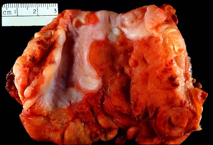
This specimen shows a PTLD which arose in the esophagus of this patient.
The white region represents mucosa of the esophagus which is partially eroded by
underlying tumor. The opposite end of the specimen consists of proximal stomach.
In this case, the tumor involved the entire esophageal wall and remained localized for
a long period of time prior to section.
Please mail comments, corrections or suggestions to the
TPIS administration at the UPMC.