


|
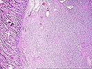
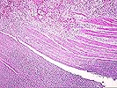
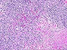
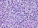
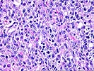
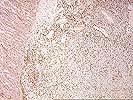
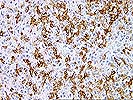

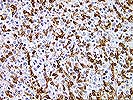
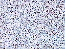
Neoplastic PTLD. Case 5. Gastrointestinal PTLD This 57 year old male underwent liver transplantation for hepatitis C-related liver disease. Six months later he presented with abdominal pain and was found to have multiple lesions involving the stomach and small bowel. Eight separate lesions were resected, of which one representative PTLD is shown here. Tumors involved the stomach and small bowel and were not seen in mesenteric nodes. Immunosuppression was modulated following surgery and the patient was alive without tumor at last followup, approximately four years later. By that time he had developed mild chronic bile duct injury of the original allograft. The H&E slides (1-3) show a lymphoid tumor which is mainly based in the submucosa and extends through and partially destroys the muscularis propria. There is a predominant population of large lymphoid cells (photographs 4-5). No convincing lymphoepithelial lesions are seen in this particular tumor. The section stained for CD20 (L26) (photographs 6-7) shows a mottled distribution of B cells throughout the tumor. Part of the reason for the irregular distribution of B cells in this tumor is that a large number of CD3-positive T cells are also present (photographs 8-9). Many of these cells have small nuclei, but some do not. Therefore, on a standard H&E section some of the "polymorphism" of these tumors may represent infiltration by nontumor cells. The EBER stain also shows an irregular distribution of EBV-positive cells within the tumor (photograph 10). The case was originally diagnosed simply as a PTLD. Some areas were polymorphic and others monomorphic, highlighting the difficulties inherent in applying a dichotomous terminology to a continuous variable. In current terminology the tumor would be considered PTLD, monomorphic, diffuse large B cell lymphoma.
Note: Click on the thumbnails to view the microscopy of this case- the "up" arrow will take you to the top of the page to see them again. Click on the back arrow to return to the previous case. Since this is the last case in this series, the forward button will take you back to a listing of all neoplastic PTLD cases. Please mail comments, corrections or suggestions to the TPIS administration at the UPMC.
If you have more questions, you can always email TPIS Administration. |
||||