


|
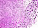
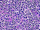
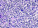
Neoplastic PTLD. Case 1 Multicentric large cell (lymphomatous) PTLD of the gastrointestinal tract This series of photomicrographs is from one of several gut tumors which arose 4 ½ months after kidney transplant in a 30 year old male under Cyclsoporine/steroid immunosuppression. He presented with an ileal perforation and underwent bowel resection. Immunosuppression was discontinued and the patient rejected his kidney and underwent allograft nephrectomy 2 weeks later. Bone marrow biopsy was negative for tumor. He was discharged and returned with melena, requiring 8 units of red cells. On barium enema examination, a lymphoid follicular pattern was seen and was considered to represent lymphoma. He underwent 3 cycles of chemotherapy with Cytoxan, Oncovin, Adriamycin and Prednisone. Repeat CT scan 4 months after chemotherapy showed no residual adenopathy. He was well for at least 5 years following diagnosis. The slides show a diffuse proliferation of mononuclear cells, many of which resemble centroblasts or immunoblasts. Lesser numbers of smaller, centrocytic or centrocytoid cells are seen and in some cases the immunoblasts contain eccentric nuclei, suggestive of early plasmacytic differentiation. Some immunoblasts are atypical, in that they are multilobated or contain deep nuclear invaginations. These cells have a tendency to occur near small foci of necrosis. The necrotic areas themselves have features of apoptosis. Finally, scattered small lymphocytes are observed. The overall appearance is that of a lymphoma with limited heterogeneity, ie, minimal polymorphism. This was originally reported as a minimally polymorphic PTLD. In current classification, it would be considered a monomorphic (lymphomatous) PTLD, immunoblastic or large cell lymphoma type. The original term monomorphic was confined to tumors with extreme homogeneity, but this has been expanded to include cases in which there is a predominant neoplastic appearing cell population. Thus, some cases which would have previously been considered polymorphic are presently classified as monomorphic PTLDs and subcategorized by lymphoma type. Southern blot showed a monoclonal B cell population in at least one tumor. These tumors have a very rapid growth rate and perforation is a common presenting sign.
Note: Click on the thumbnails to view the microscopy of this case- the "up" arrow will take you to the top of the page to see them again. Click on the forward button to go to the next case. Click on the back arrow to return to a listing of all neoplastic PTLD cases. Please mail comments, corrections or suggestions to the TPIS administration at the UPMC.
If you have more questions, you can always email TPIS Administration. |
||||