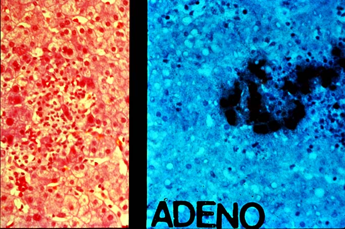
Figure 1.
The left side of the photomicrograph shows the
typical routine histopathological appearance of
adenoviral hepatitis. Note the poorly formed
granulomatoid collection of macrophages, intermixed
with partially viable hepatocytes. At this magnification,
it is too difficult to recognize the inclusion-bearing
cells.
The right side of the photomicrograph illustrates
positive immunohistocemical staining for viral antigens,
which confirms the diagnosis, if one is unsure about the
changes on routine light microscopy.
Please mail comments, corrections or suggestions to the
TPIS administration at the UPMC.