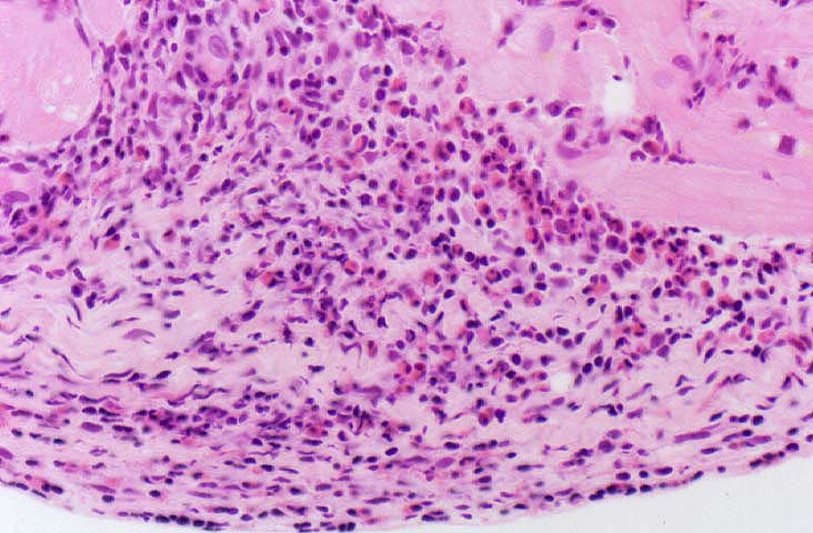
Figure 9.
A higher magnification of figure 8, illustrates the differences between
the endocardial inflammation of acute rejection and Quilty lesions.
Note the composition of the infiltrate in this case, which contains a
significant number of eosinophils and blastic lymphocytes. Quilty lesions
on the other hand, are usually composed on small lymphocytes and there
are small blood vessel with prominent endothelium amidst the
inflammation of a Quilty lesion.
Please mail comments, corrections or suggestions to the
TPIS
administration at the UPMC.