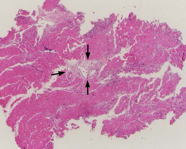
Figure 1.
This is a typical example of ischemia/reperfusion
injury in a cardiac allograft biopsy obtained 10 days after
transplantation. The most important distinguishing features are
areas of myocyte dropout (arrows) which are not associated
with significant mononuclear inflammation. A variable number
of neutrophils and pigmented macrophages may be present.
Please mail comments, corrections or suggestions to the
TPIS
administration at the UPMC.